Recognizing skin cancer symptoms using model based imaging

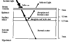
When a quality skin model is constructed – recognizing skin cancer symptoms can be more comfortable as many factors indicate the threat of skin cancer. Of course, this can’t give 100% results, as there are many shortcomings connected with skin lesion variety and interpretation errors. But some guides may help. Three main factors can indicate the risk of skin cancer. Recognizing skin cancer symptoms can be based on them. They are: Melanin presence in the papillary dermis; The thickness of papillary dermis; Blood behaviour around the lesion and inside it. The first important factor is the melanin present in the dermis. This is the main factor in recognizing skin cancer symptoms. If melanin spread in the papillary dermis or even dermis, this is a significant probability of being skin cancer symptoms, but not always. Several subfactors in this issue, like melanin spreading figure, depth, and melanin density within this shape. If there are more irregularities in the spreading area, there are more risks. Another factor in recognizing skin cancer symptoms is papillary layer thickness. In not going into deep too much there can be said, that the thinner this layer, the more significant risk.