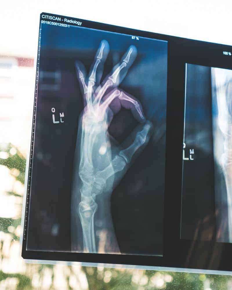
Category: Health
Recognizing skin cancer symptoms using model based imaging

When a quality skin model is constructed – recognizing skin cancer symptoms can be more comfortable as many factors indicate the threat of skin cancer. Of course, this can’t give 100% results, as there are many shortcomings connected with skin lesion variety and interpretation errors. But some guides may help. Three main factors can indicate the risk of skin cancer. Recognizing skin cancer symptoms can be based on them. They are: Melanin presence in the papillary dermis; The thickness of papillary dermis; Blood behaviour around the lesion and inside it. The first important factor is the melanin present in the dermis. This is the main factor in recognizing skin cancer symptoms. If melanin spread in the papillary dermis or even dermis, this is a significant probability of being skin cancer symptoms, but not always. Several subfactors in this issue, like melanin spreading figure, depth, and melanin density within this shape. If there are more irregularities in the spreading area, there are more risks. Another factor in recognizing skin cancer symptoms is papillary layer thickness. In not going into deep too much there can be said, that the thinner this layer, the more significant risk.
DullRazor – digital skin hair shaver

DullRazor uses image processing techniques to analyze and segments skin areas with dark hair. This program removes dark hair form images and makes skin lesion images clean to further processing. From: To: Many skin images contain various numbers of hairs. Other skin segmentation programs may mislead because of hairs – especially dark ones. One solution can be shaving skin before taking pictures of it. But shaving of skin adds more time to processing, and this is uncomfortable and, in some cases, unaesthetic. Hence, a software approach for dark, thick hair removal from skin images are needed.
Skin Cancer causing factors

Substance Where this can be found How to avoid Arsenic Pesticides, wood preservatives, alloy additive non-ferrous metals. Use protective clothing when working with arsenic substances Creosote Wood preservative Use protective clothing when working with creosote substances Ionizing radiation Ionizing radiation is specific industrial sterilization sources Limit exposure if possible. Wear a dosimeter while working with radiation. Sunlight Summer, and when on a sun holiday. Avoid strong sunlight, especially at midday. Wear protective clothing to protect your skin. Cover exposed skin with sunscreen of factor 15 or higher. Tar Coal tar Use protective clothing Glutaraldehyde Glutaraldehyde is used as a disinfectant. It is also can be found in X-ray films. Use protective clothing when dealing with glutaraldehyde. Work only in well-ventilated areas. Soot Black particles of carbon, produced by incomplete combustion of coal, oil, wood, or other fuels Use protective clothing Pitch It is made by the destructive distillation of wood or coal tar Use protective clothing Asphalt Sticky, black and highly viscous liquid or semi-solid that is present in most crude petroleum and in some natural deposits Use protective clothing Paraffin wax A member of the alkenes series Use Gloves Smoking Smoking cigarettes increase your risk of cell carcinoma…
Healthy skin reflectance model

This pilot study is intended to investigate possibilities of skin nevus imaging using digital still image camera. The main objective is to develop a dermatology image interpretation method, which enables the looking on the skin lesions and nevus from the optical background of skin colouration. Kubelka-Munk calculation method for light transport and reflection from multilayered, complex media is applied to model light reflection spectra of skin. Calculation of model shows that red, green, blue and infrared colours lighting is satisfactory to access distribution of comparative estimates of the following skin parameters: volume fraction of melanin in the epidermal layer, the volume fraction of haemoglobin in the dermal layer, presence of dermal melanin and thickness of the papillary layer. Performance of image processing method on fourteen samples of images of common melanocytic nevi, dysplasia melanocytic nevi, Spitz nevus, thrombotic hemangioma and surrounding healthy skin were made. Skin spectral properties Understanding how light interacts with skin can assist in designing physics-based dermatological image processing. The key is understanding how light interacts with skin tissue. The skin consists of different layers with different spectral properties. Fig 1. Skin model and its physical view When incident light is applied to skin layer, it is…
Review on skin lesion imaging, analysis and automatic classification

The goal of any imaging methodology used in dermatology is to diagnose melanoma in the early stages because it depends on treatment effectiveness. Investigations show that early diagnosis is more than 90% curable and late is less than 50% [1]. The diagnosis and successful treatment are often supplemented with permanent monitoring of suspicious skin lesions. The doctor’s diagnosis is reliable, but this procedure takes lots of time, effort. These routines can be automated. It could save lots of doctor’s time and could help to diagnose more accurately. Besides using computerized means, there is an excellent opportunity to store information with diagnostic information to use it for further investigations or create new methods of diagnosis. Skin lesion imaging methods We found that there are several various imaging methods of skin lesions [2]. The simplest skin visualization method is photography. This method gives only the top layer of the skin image. To get a deeper layer image, there is oil immersion used. It reduces reflections of the surface and brightens the image of the epidermis – the second skin layer.
Skin lesion boundary tracing algorithm

I found Matlab to be a convenient tool that easily traces the boundaries of objects in a picture. So I adopted it to skin lesions. This can be used to automatically detect skin irregularities and calculate lesion properties like the asymmetry of shape, or border irregularities, which can help detect melanoma. There are numerous investigations done, so I only put a few examples of how it looks. I will give you my source code so that you can try it on your own. Look at my results: 1) And it also finds the center of mass:
Skin imaging methods for melanoma diagnosis

There are many skin image capture methodologies developed and used. Here is a short review of them: Dermatoscopic photography The deepest layer of skin can be reached – Papillary dermis Resolution – depends on the optical system View of skin – Horizontal The main disadvantage is reflections of light from skin surface – stratum cornea.