
Magnetic resonance imaging (MRI) is a common medical procedure requested by doctors for patients they suspect as having a serious medical condition. Apart from checking the brain and the spinal cord, MRIs can generate a visual representation of your internal organs.
With this medical procedure, doctors could provide better diagnoses and suggest appropriate ways to move forward. Your physician may suggest further tests or immediately proceed to work on a treatment plan in some cases.
Whether you’ve just been told to undergo an MRI scan and don’t know what to expect, or want to know more about this advanced medical equipment, read on to learn the basics of MRI: what it is, what its main uses are, and its limitations.
What Is An MRI And How Does It Work?
Magnetic resonance imaging (MRI) is a medical test that harnesses the power of magnets, radio waves, and computer equipment to generate detailed images inside your body. An MRI scan is typically requested after other medical tests have failed to provide information for appropriate medical diagnosis. MRI’s accuracy allows it to distinguish between normal and damaged or abnormal tissues. It works even better than an X-ray, CT scan, and ultrasound procedure.
MRI scanners are said to be relatively safer compared to other machines such as X-rays and computed tomography (CT) scans, which are believed to utilize potentially damaging ionizing radiation in its processes.
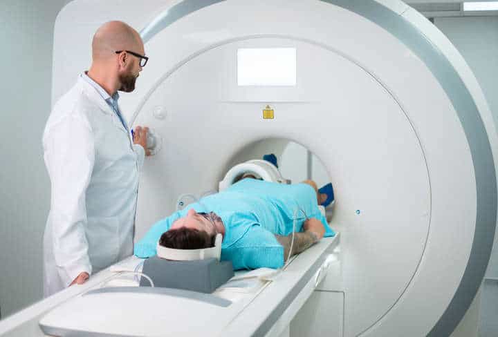
1. Traditional MRI Scanning Machine
This full body MRI scan machine features a tunnel-like tube where the patient is placed. The tube is covered by a circular magnet from which a strong magnetic field emanates, arranging the protons of hydrogen atoms that further interact with radio waves. As the radio waves and magnetic field interact, it stirs the protons in your body, generating a signal that’s caught by the MRI detector. The data is passed on to the computer, which processes the information and produces the accurate image displayed in the monitor where a radiologist and your physician sit.
The machine produces high-quality and detailed images of a specific body part under examination. This makes detecting structural changes in your body highly visible. If a more accurate image is needed, an element called gadolinium is injected. This contrast agent assists physicians in identifying inflammation, tumors, and blood clots because it helps the MRI scanner produce clearer and overall better images.
2. Short-bore MRI Scanning Machine
Apart from the traditional MRI scanning machine, scanning can be done even without placing the patient’s whole body inside the tube. For instance, in a short-bore system, only a specific portion of your body is scanned by the machine. As an alternative MRI scanning method, the patient only has to insert the body part that needs to be examined. The rest of the patient’s body stays outside of the scanner.
3. Open MRI Scanning Machine
Another MRI machine doesn’t look like a tunnel, perfect for claustrophobic and overweight patients. Aptly called an open MRI, it doesn’t have walls on either side. Because it produces a weaker magnetic field and radio waves, the machine’s images are not as clear as those produced by an enclosed MRI.
What Are The Uses of MRI?

These advanced electronic imaging tools can be used in many ways. For one, it can validate your specific health condition. That being said, it’s a key medical tool to achieve correct physician diagnosis. With proper diagnosis, your doctor will be able to recommend the appropriate medical care and treatment for your condition. In some cases, your healthcare professional can also use the MRI image to determine your body’s response to the treatment.
The MRI scanner can be used for a lot of applications. It can generate images from the following body parts:
- Internal organs within the chest and abdominal area include the heart, liver, kidneys, adrenal glands, and other organs. As such, it can confirm the lung diseases and abnormalities in the said body parts. Congenital heart disease and the overall structure of the heart, including the damaging effects of a heart attack, are also reflected in the images.
- Reproductive and internal organs in the pelvic area include the uterus, ovaries, prostate gland, and bladder.
- Your blood vessels and lymph nodes, which may be useful in detecting certain types of cancer. Blood vessel issues and inflammation, as in the case of vasculitis, may also be detected and blocked blood vessels.
- MRI machines can detect tumors in almost any part of the body, as they can produce images from the soft tissues of your internal organs and the nervous system.
- It can detect inflammation and inflammatory diseases, for instance, Crohn’s disease.
- An MRI scanning can detect a fetus in the womb and check for abnormalities in a woman’s nipple discharge and breast cancer.
- In the brain and spinal cord, an MRI scanning can check for damages, injuries, and problems in the eye and the inner ear. Health conditions such as cancer and multiple sclerosis can also be confirmed through this medical equipment. Brain aneurysms can also be recognized.
- Neurosurgeons rely on MRI scanning to ensure the soundness of the spinal cord following a traumatic injury.
- In scanning your bones and joints, an MRI scanner can locate cancer cells, bone infection, and damages, problems with the spinal discs, nerve pinching in the lower and upper back.
- A functional MRI (fMRI) can map brain activities to detect potential problems. This special machine monitors the blood flow in the brain to see the areas that become functional when you perform particular activities. This machine can be used for patients who are candidates for surgeries to treat epilepsy and tumors.
MRI scans are likewise often used by surgeons prior to surgery in order to properly locate the problem areas or regions of concern.
What Are The Limitations of MRI?
The key to generating high-quality and accurate images relies on the patient’s ability to keep still while being scanned. Another contributing factor is following the instructions when to inhale and exhale while the images are being recorded. Even an irregular heartbeat can impact imaging the procedure, being that some machines use the heart activity to time each capture. Also, some MRI machines have size and weight limitations.
Here are additional impediments to producing clear images and few additional limitations of an MRI machine:
- Metallic objects
- Excessive patient movement
- Incorrect breathing
- Bowel motion for pelvic and abdominal scans
- Edema and cancer fluids may look similar when scanned.
Although relatively safe, women in their first trimester of pregnancy should avoid being scanned except in cases of medical necessity. In the same vein, patients with pacemakers, metallic ear implants, bullet fragments should best avoid MRI procedure. Individuals undergoing chemotherapy and insulin pumps should avoid it, as well.