There are many skin image capture methodologies developed and used. Here is a short review of them:
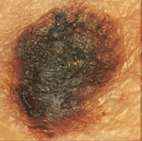
Dermatoscopic photography
- The deepest layer of skin can be reached – Papillary dermis
- Resolution – depends on the optical system
- View of skin – Horizontal
The main disadvantage is reflections of light from skin surface – stratum cornea.
Dermatoscopic oil immersion photography
- The deepest layer of skin can be reached – Papillary dermis
- Resolution – depends on the optical system
- View of skin – Horizontal
Reflections of light from skin surface are smaller because of oil used between camera optics and skin.
Fluid free dermatoscopic photography, polarized light
This method gives similar results to using oil immersion. It is a cleaner image taking way because sin is kept dry and there is no wet contact with the camera.
Trimodal light source
- The deepest layer of skin can be reached – Papillary dermis
- Resolution – depends on the optical system
- View of skin – Horizontal
This is a unique method of light directing to deeper skin layers avoiding passing the lesion surface. It gives outstanding results of skin deeper layer imaging.
CSLM
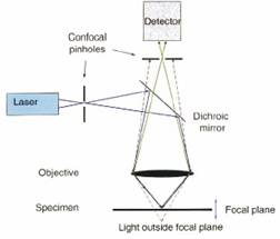
CLSM – laser scanning microscope system. Their laser beam is focused on the particular skin layer and scanned 2D image in this deep. This method is handy when taking deeper layers of skin. The deep can be from 2 to 300ukm.
Multispectral dermatoscopy
This method is described in earlier articles. There is multispectral light used to take separate pictures. The calculation is used to evaluate the pigmentation of different skin layers. This is a progressive methodology in diagnosis.
Optical Coherence Tomography
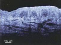
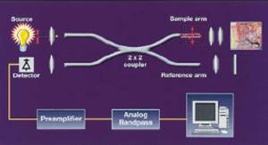
There is interference of two different light sources. One is directed to the skin, and other is a reference to determine the depth. This method is more advanced and not suitable for screening. The depth of imaging is in the range between 15 and 2 μm.
High-Resolution Ultrasonic Imaging
There is high-frequency ultrasound used to determine lesion boundaries before surgery. In early skin cancer detection, this method hardly can be used, as the difference between normal and suspicious regions are minimal. But combining Ultrasound and let us say multispectral photography can show good results.







Wow! Informative Webpage.
another great article